
国際的に権威ある学術誌に掲載
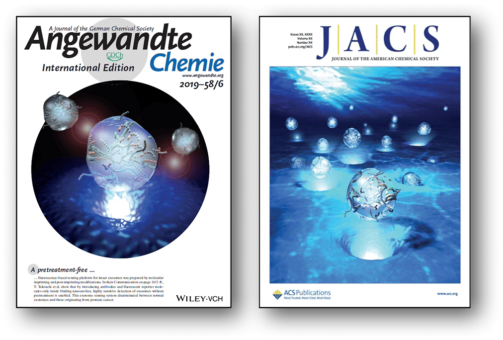
神戸大学の研究成果であるTearExo法は、
国際的に権威ある学術誌に掲載されています。
Angewandte Chemie International Edition(ドイツ化学系トップジャーナル)、Journal of the American Chemical Society(米国化学系トップジャーナル)にて、ジャーナルカバーイメージとともに掲載されました。

Research

神戸大学の研究成果に基づく、涙液中エクソソームを簡便、高感度に測定する革新的な技術
About
TearExoの涙液検査は、従来の乳がん検診に代わるものではありません。
定期的な乳がん検診の重要性は変わりません。
この検査は、検診と検診の間に「今の自分のリスクはどうだろう」と確かめたいとき、乳がんについて考えるきっかけとなる新しい選択肢です。
涙液中に含まれる「細胞外小胞(エクソソーム)」という物質に着目。がん細胞と正常細胞では異なる性質を持つことが知られています。
神戸大学の研究成果に基づき、涙液中のエクソソームを高感度で測定できる技術を開発。従来の測定法の1,000倍の感度を実現。
Results
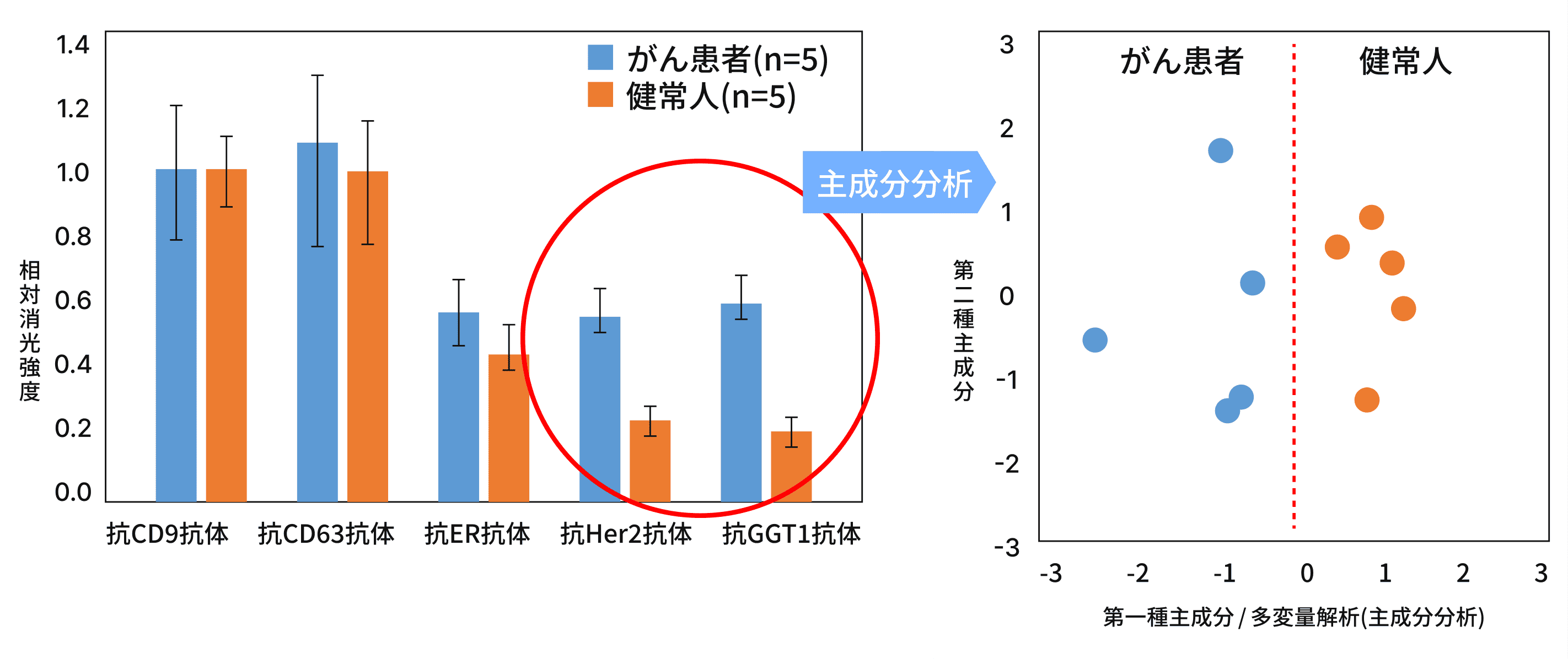

涙液中のエクソソームを測定することで、乳がん患者とがんがない方のエクソソームに違いがあることが確認されました。つまり、涙液による乳がんリスク判別の可能性が示されました。
乳房全摘手術を受けた患者さんの手術前と手術後で、涙液中のエクソソームが変化することも確認されています。
手術後には健常な方と同様のエクソソームが検出されたことから、将来的には薬物療法の効果確認や術後の経過観察、再発リスクのチェックなど、様々な場面での活用可能性も考えられます。
Method

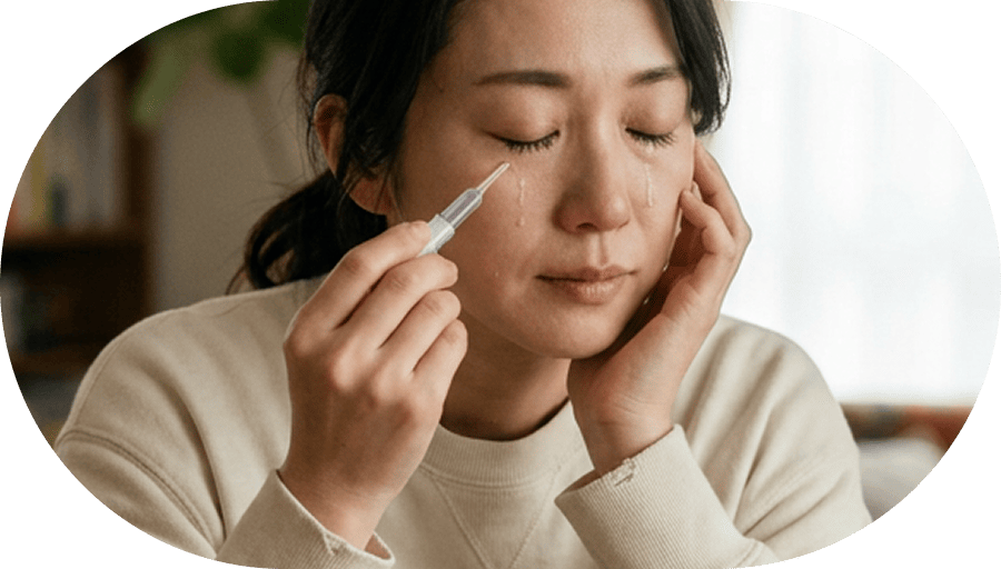
ドライアイの検査でも使われる「シルマー試験紙」という、小さくて薄い短冊状のろ紙を目じりに置き、数分間目を閉じて涙をしみこませます。
採取した試験紙は専用の溶液に浸してエクソソームを回収し、自動分析装置で測定します。


将来的には、新しく開発する予定のさらに非侵襲的な涙液採取法への転換も検討しています。
乳がんについて考え、行動するきっかけを、もっと身近にしたいと考えています。
Method
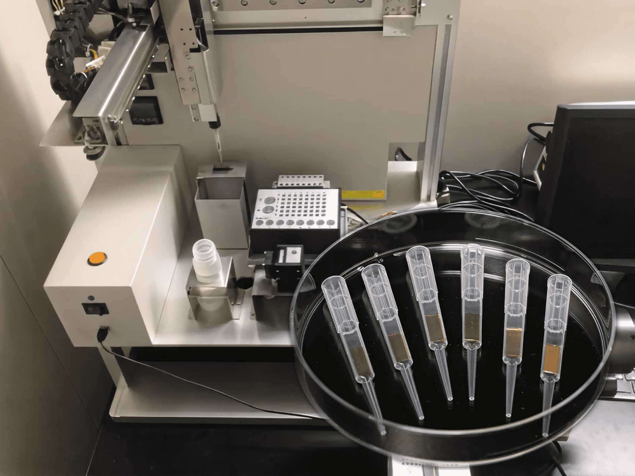
専用のセンシングチップを準備します。
シルマー試験紙で採取した涙液を抽出します。
センシングチップが装着された分析装置で涙液成分を分析し、結果が出力されます。
Original Article
神戸大学の研究成果であるTearExo法は、
国際的に権威ある学術誌に掲載されています。
Angewandte Chemie International Edition(ドイツ化学系トップジャーナル)、Journal of the American Chemical Society(米国化学系トップジャーナル)にて、ジャーナルカバーイメージとともに掲載されました。
Mori, K., Hirase, M., Morishige, T., Takano, E., Sunayama, H., Kitayama, Y., Inubushi, S., Sasaki, R., Yashiro, M., Takeuchi, T. A pretreatment-free, polymer-based platform prepared by molecular imprinting and post-imprinting modifications for sensing intact exosomes, Angew. Chem. Int. Ed. 2019, 58, 1612-1615. (DOI: 10.1002/anie.201811142)
Takeuchi, T., Mori, K., Sunayama, H., Takano, E., Kitayama, Y., Shimizu, T., Hirose, Y., Inubushi, S., Sasaki, R., Tanino, H. Antibody-conjugated signaling nanocavities fabricated by dynamic molding for detecting cancers using small extracellular vesicle markers from tears, J. Am. Chem. Soc. 2020, 142, 6617-6624. (DOI: 10.1021/jacs.9b13874)